Welcome to a journey through the vital elements of Picture Archiving and Communication Systems (PACS). As healthcare continues to embrace digital transformations, understanding PACS components becomes essential for technologists and anyone involved in patient care.
This guide breaks down the core components of PACS, highlighting their roles and how they collectively enhance the management of medical images. Each element is pivotal in streamlining diagnostics and treatment plans, from the capturing devices that digitize images to the sophisticated networks that share them.
Whether you’re a seasoned radiologist or a healthcare administrator aiming to optimize your facility's imaging capabilities, this detailed exploration will equip you with the knowledge to leverage PACS technology effectively, ensuring efficient and comprehensive patient care.
Join us as we unravel how each part of PACS contributes to the bigger picture of healthcare excellence.
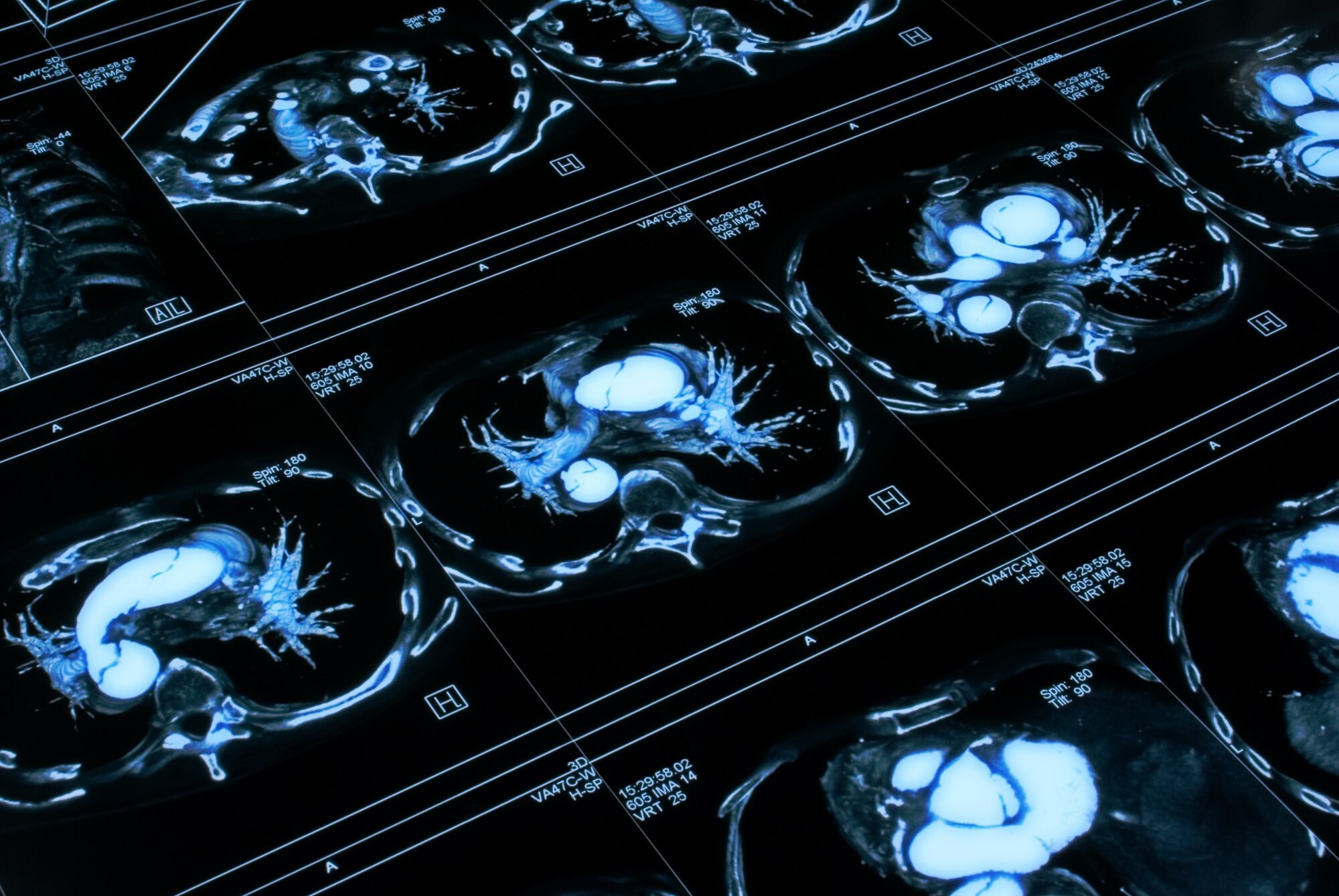
Imaging modalities are the cornerstone of diagnostic medical imaging. Essentially, these technologies capture human body images to aid diagnosis and treatment.
In Picture Archiving and Communication Systems (PACS), these modalities serve as the primary data source, feeding detailed, essential medical images into the system.
The role of these modalities in PACS is critical—they capture images and tag them with metadata that facilitates efficient categorization and retrieval within the PACS architecture.
Several types of imaging modalities are integral to modern medicine:
• Magnetic Resonance Imaging (mri): Uses strong magnetic fields and radio waves to produce detailed images of the organs and tissues within the body.
• Computed Tomography (ct) Scans: Utilizes X-rays to create comprehensive cross-sectional views of the body, providing more detail than regular X-ray exams.
• X-rays: One of the oldest and most frequently used forms of medical imaging, essential for diagnosing conditions in the chest and bones.
Each modality is selected based on the specific diagnostic needs and the body part being examined. For instance, MRIs are particularly useful for imaging soft tissues, while CT scans are often preferred for quicker, detailed examinations of internal organs, bone, soft tissue, and blood vessels.
Standard protocols, predominantly DICOM (Digital Imaging and Communications in Medicine), facilitate the integration of these modalities with PACS. DICOM enables the seamless transmission of medical images and their associated information across different systems and devices within healthcare settings.
When any of these modalities captures an image, it is automatically converted into a DICOM format, which includes vital patient data and image specifications. This standardization ensures that the images can be easily accessed, viewed, and analyzed across various PACS workstations regardless of the origin or type of imaging modality.
This integration is not just about storage; it also enhances image accessibility. Radiologists and physicians can retrieve and review these images from PACS, often from remote locations, enabling timely and informed medical decisions. This connectivity exemplifies how crucial imaging modalities are in the overarching medical diagnostics and patient care framework facilitated by advanced PACS technologies.
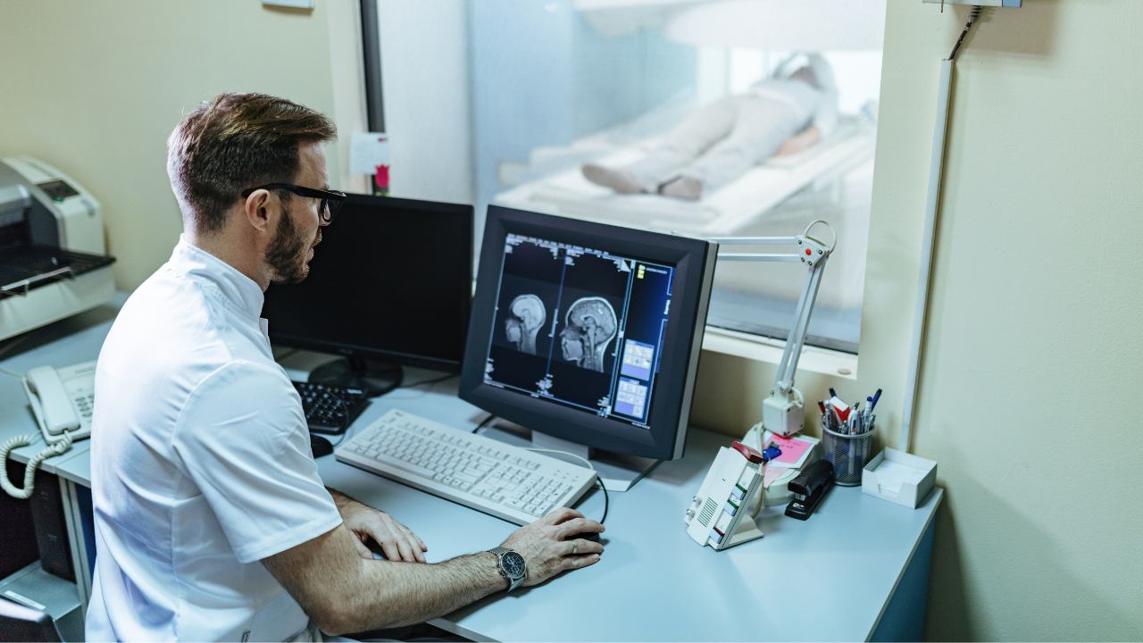
Acquisition devices are fundamental to the intricate PACS (Picture Archiving and Communication System) system.
These devices are specifically designed to capture medical images from various imaging modalities and are integral to the seamless operation of PACS. They bridge raw imaging data and digital storage, ensuring that every captured image is accurately rendered and stored.
Acquisition devices are tasked with a critical function: they capture images and convert them into a digital format that PACS can process. This involves digitizing analog signals (in cases where older imaging technology is used) and ensuring that digital images are formatted correctly.
These devices embed essential metadata into each image, such as patient ID, date of acquisition, and specific details pertinent to the study. This metadata is crucial for organizing and retrieving images efficiently within the PACS.
Moreover, these devices often perform preliminary image processing to enhance the quality of the photos before they are stored and viewed on PACS, thus ensuring that clinicians have access to the highest quality images for diagnosis.
The variety of acquisition devices corresponds to the range of imaging modalities used in medical diagnostics. For instance:
• Digital Radiography Panels: These panels capture images digitally and are known for swiftly delivering high-resolution images in X-ray systems.
• Ct Scanners: These complex machines include built-in acquisition devices that handle the enormous data output from CT scans, processing and converting it into digital formats easily managed by PACS.
• Ultrasound Imaging Systems: These utilize advanced transducers that act as acquisition devices, converting sound waves into visible images immediately available for integration into PACS.
Each type of acquisition device is tailored to meet the demands of the specific imaging technique it supports, ensuring optimal compatibility and performance. From the rapid image processing requirements of an MRI scanner to the high-resolution needs of digital mammography, these devices are equipped to handle a range of functionalities, making them indispensable in the digital imaging ecosystem.
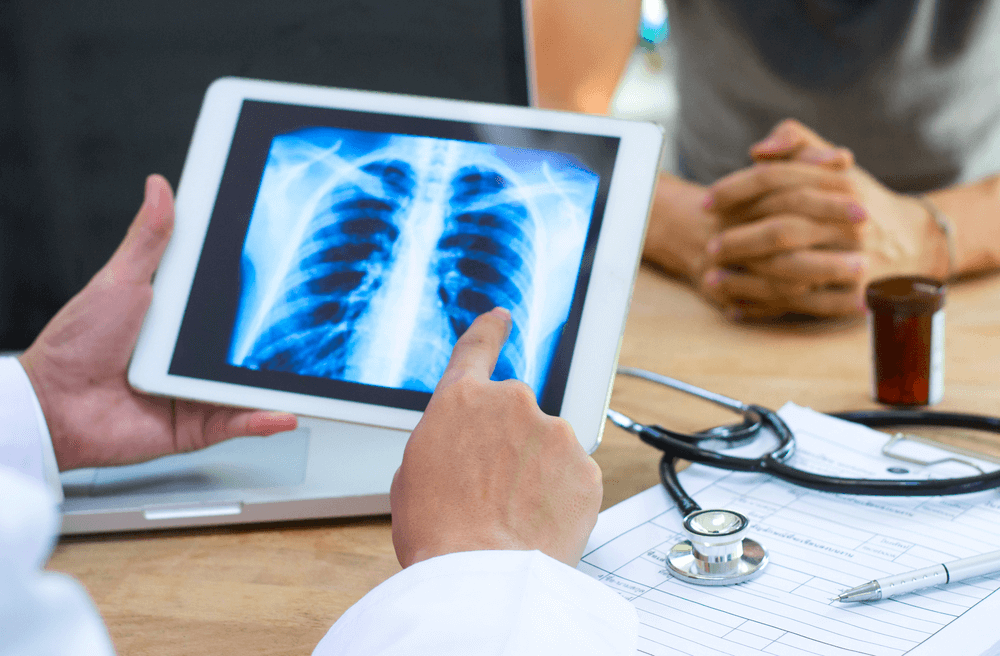
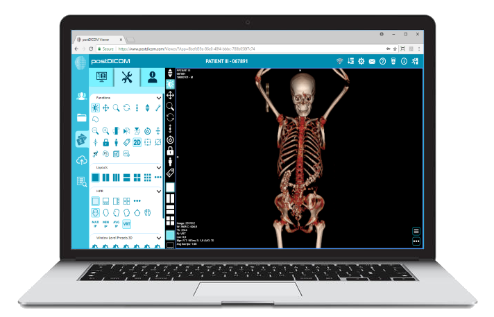
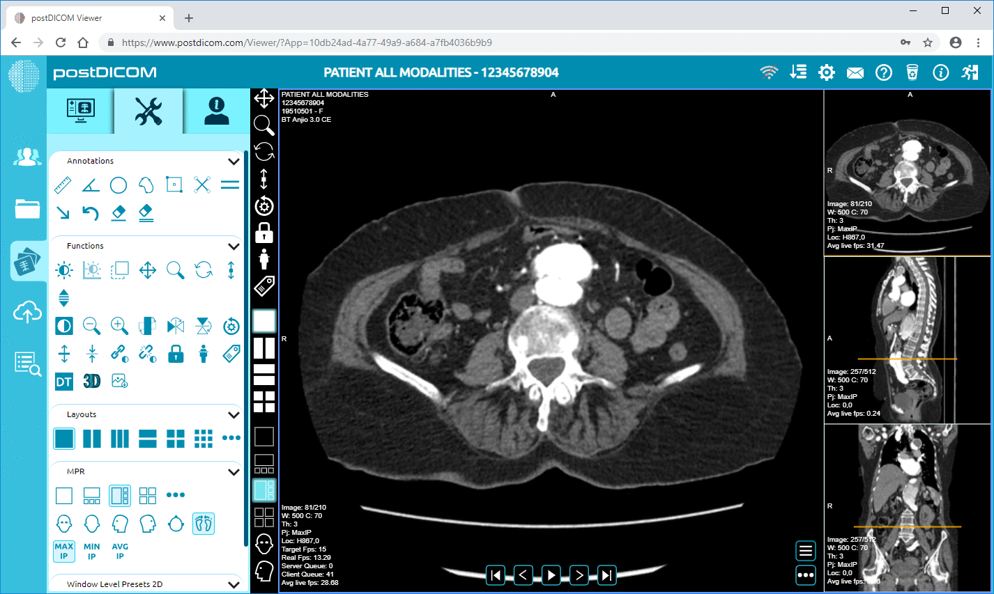
In the PACS ecosystem, workstations serve as the critical interface where medical images are handled digitally.
These powerful computers are tailored to meet the demanding needs of medical imaging, enabling radiologists and medical professionals to view, analyze, and manipulate digital images efficiently. Their primary role is to provide a reliable, high-performance platform that supports the complex software needed for detailed medical image assessments.
The viewing software loaded onto PACS workstations is robust and equipped with tools to enhance the diagnostic process. Key features include:
• Advanced Image Processingallows Users To Adjust Brightness, Contrast, And Zoom On Images To View Fine Details More Clearly, Which Is Essential For Accurate Diagnosis.
• Annotations And Measurements: Software tools enable the addition of markers, notes, and measurements directly on the images, which are crucial for surgical planning and tracking changes over time.
• 3d Reconstruction: Some advanced PACS software can reconstruct two-dimensional images into three-dimensional models, providing a more comprehensive view of the studied anatomical structure.
These features are crucial for making precise diagnoses and are particularly beneficial in specialties such as orthopedics, where detailed imagery can significantly impact treatment plans.
The effectiveness of a PACS workstation is greatly influenced by the design of its user interface. A user-friendly interface simplifies the complexity inherent in medical imaging, enabling medical staff to navigate through functions easily and efficiently. The ideal interface should:
• Minimize Clicks: Reduce the number of interactions needed to perform everyday tasks, speeding up the workflow.
• Intuitive Layout: Arrange tools and menus logically so that new users can learn the system quickly and experienced users can work efficiently.
• Customizability: Allow users to adjust the layout and settings to fit their personal preferences and the specific needs of their medical specialty, which can vary significantly from radiology to cardiology.
These aspects of the interface design contribute to operational efficiency and reduce users' cognitive load, allowing them to focus more on diagnostic tasks rather than on navigating the software.
Well-designed workstations and software are not just tools but active participants in the diagnostic process. They enhance the capabilities of healthcare providers and ultimately improve patient outcomes through better, faster diagnostic services.
As technology evolves, these systems continue to adapt, incorporating more intuitive designs and features that anticipate the needs of modern medical environments.
Archive servers form the backbone of a PACS system, providing robust storage solutions for the vast amounts of imaging data that healthcare facilities generate daily.
These servers are not just repositories; they organize and manage medical images and associated diagnostic data effectively, ensuring that every data byte can be retrieved and reviewed when needed. This is crucial in environments where historical data comparison is necessary for accurate diagnosis.
The technology powering these servers is designed for both efficiency and reliability:
• Data Compression: To manage massive data volumes, archive servers use sophisticated compression algorithms that reduce the storage space needed per image without losing image quality, which is crucial for detailed medical analysis.
• Redundancy: These systems often employ redundancy techniques such as RAID (Redundant Array of Independent Disks) configurations, which ensure that even if one disk fails, there is no data loss. This redundancy is essential for maintaining data integrity and continuous access to medical records.
Security is paramount in medical imaging due to the sensitive nature of the data stored. Archive servers are equipped with multiple layers of security measures:
• Encryption: Data at rest and in transit are encrypted using advanced encryption standards, ensuring unauthorized individuals cannot access or interpret the data.
• Access Controls: Rigorous access controls are implemented, requiring user authentication and maintaining detailed access logs to track who accessed data and when. This helps maintain compliance with regulations like HIPAA, which demand strict handling and confidentiality of patient information.
• Regular Audits: Security protocols include regular audits to identify and rectify potential vulnerabilities, ensuring the system’s defenses remain robust against evolving threats.
These servers are more than mere storage facilities; they are a critical asset in the digital infrastructure of modern healthcare, providing not just storage but also security and data management solutions that uphold the integrity and accessibility of vital medical data. As technology progresses, the capabilities of these servers continue to evolve, offering more advanced features that enhance the overall efficiency and security of PACS systems.
The backbone of any Picture Archiving and Communication System (PACS) is its communication network, which handles the hefty task of transmitting imaging data across various points in a healthcare setting.
The smooth operation of a PACS largely depends on these networks, which connect different system components, such as scanners, archive servers, and workstations, enabling real-time data access and sharing.
The efficiency of a PACS network hinges on several key components:
• Routers And Switches: These devices direct data traffic efficiently across the network, ensuring that imaging data reaches its intended destination swiftly and without bottlenecks. They are crucial in maintaining the flow of large image files, typical in medical imaging.
• Firewalls: As gatekeepers, firewalls provide a critical security layer, controlling incoming and outgoing network traffic based on security rules. This is vital in protecting sensitive medical data from unauthorized access.
Given the sensitive nature of medical data, communication networks within PACS must adhere to stringent security and compliance standards:
• Data Encryption: To safeguard privacy and integrity, data transmitted over the network is encrypted, making it unreadable to anyone without authorized access.
• Compliance Standards: Networks must comply with health information privacy laws, such as HIPAA in the U.S., which sets standards for protecting health information. Compliance ensures that the network is not only secure but also legally sound.
• Regular Security Assessments: To keep up with evolving threats, networks undergo regular security assessments and updates. This proactive approach helps identify vulnerabilities and apply necessary patches or security enhancements.
The robustness of communication networks within PACS is about maintaining operational efficiency and ensuring that every component functions seamlessly in a secure and compliant environment.
As technology advances, these networks evolve, incorporating newer technologies that promise even greater efficiency and security, making them indispensable in the modern healthcare landscape.
A pivotal aspect of modern healthcare IT infrastructure is the seamless integration of PACS with Radiology Information Systems (RIS) and Electronic Health Records (EHR). This integration allows for a streamlined flow of information across different systems, enhancing healthcare providers' capability to access and utilize data efficiently.
PACS interfaces with RIS to manage imaging orders and track radiology reports, linking diagnostic imaging data directly to the patient’s broader medical record managed within the EHR system.
This connectivity ensures that when a radiologist captures and uploads an image to PACS, the image can be viewed alongside the patient’s medical history, laboratory results, and other diagnostic information stored in the EHR.
 - Presented by PostDICOM.jpg)
• Improved Data Accessibility: With PACS integrated into EHR and RIS, medical images are readily accessible to all authorized healthcare providers, regardless of location. This accessibility is crucial for consultations and can significantly speed up diagnosis and treatment processes.
• Workflow Efficiency: Integration minimizes the steps required to access patient information and images. Physicians and radiologists do not need to switch between multiple systems; instead, they have a unified interface that provides all the necessary data, reducing time and potential errors.
• Enhanced Patient Care: Immediate access to complete patient records and associated images leads to better-informed decisions, more accurate diagnoses, and tailored treatment plans. This not only improves the quality of care but also enhances patient outcomes.
This integration represents a leap towards a more interconnected healthcare environment where data flows seamlessly between departments, increasing the effectiveness of medical diagnostics and patient management. As technology progresses, the depth of integration is only expected to increase, making comprehensive patient care more efficient and precise.
The components of PACS—ranging from advanced imaging modalities and acquisition devices to robust archive servers and comprehensive communication networks—form the backbone of modern medical imaging.
Integrating these systems streamlines radiological workflows and bridges the gap between various health information systems, enhancing the quality and speed of patient care.
As technology continues to evolve, so does PACS's potential to transform healthcare delivery. With ongoing advancements in AI, machine learning, and telemedicine, PACS is set to play an even more crucial role in diagnostic precision and accessibility.
This progression underscores the importance of choosing a PACS provider like PostDICOM that stays at the forefront of innovation. This ensures that healthcare facilities are equipped with the best tools to meet the demands of modern medicine.
Embracing PACS means not just adopting new technology but advancing a culture of efficiency and safety in healthcare, where every component works seamlessly to support healthcare professionals and enhance patient outcomes.
|
Cloud PACS and Online DICOM ViewerUpload DICOM images and clinical documents to PostDICOM servers. Store, view, collaborate, and share your medical imaging files. |